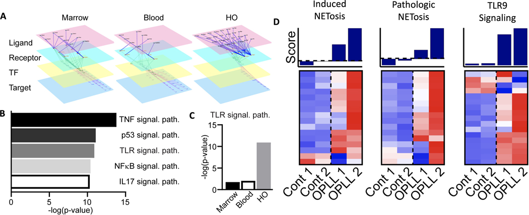
Figure 4. Upregulation of TLR and NETosis pathways in ectopic bone formation in a mouse and human model.
(A) Representative diagram of scRNA-seq based multilayer networks done by integrating intercellular pathways (ligand-receptor interactions) and intracellular subnetworks (receptor-transcription factor and transcription factor-target gene interactions) in relation to the neutrophil target. Sequential activation of receptors by their ligands, their downstream transcription factors, and the transcription factor target genes is shown. (B) Pathways upregulated in neutrophils at the HO injury site from scRNA-seq based multilayer network analysis. Higher -log(p-value) is associated with more enrichment of the pathway. (C) Inferred TLR9 pathway activation across anatomical sites. Higher-log(p-value) is associated with more enrichment of the pathway. (D) Microarray analyses of ossification of the posterior longitudinal ligament (OPLL) using gene lists for induced NETosis and pathologic NETosis, and TLR9 signaling pathways were compared in healthy controls (n=2) versus those with OPLL (n=2).

