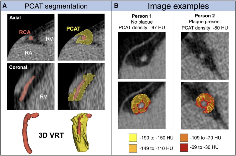
Figure 2.
Pericoronary adipose tissue on noncontrast cardiac computed tomography. A, Segmentation of PCAT surrounding the right coronary artery to measure PCAT density. B, Examples of patients with and without CAD and correspondingly higher PCAT density in the patient with CAD. 3D VRT, 3-dimensional volume rendering technique; CAD, coronary artery disease; HU, Hounsfield unit; PCAT, pericoronary adipose tissue; RA, right atrium; RCA, right coronary artery; RV, right ventricle; VRT, volume-rendering technique.

