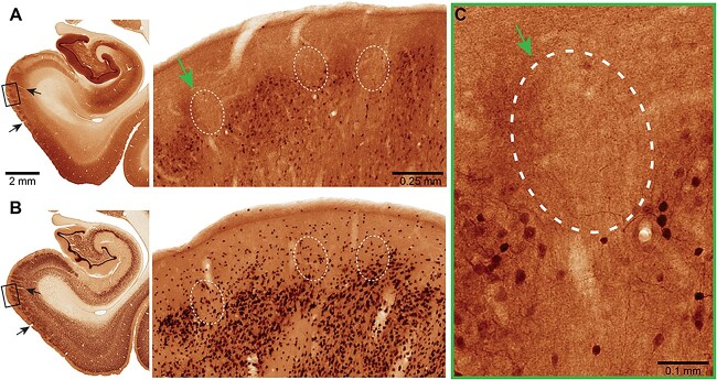
Fig. 12.
Brightfield micrographs revealing calbindin immunoreactivity in ERC. MacBrain Resource Collection 6, Brain 79, female 9.80 years old. (A) Calbindin (CB) and (B) neuronal nuclear antigen (NeuN) staining in the entorhinal cortex. The CB section is 700 μm rostral to the NeuN one and they correspond to section number 38 in each series, publicly available at https://macbraingallery.yale.edu/collection6/#B79. The black arrows point to capillaries that were followed for spatial matching. White dashed ovals in (A) show layer II areas devoid of CB+ cells and in (B) the approximate corresponding areas. Scale bars in (A) apply to (B). (C) High magnification micrograph showing areas in ERC layer II cell clusters devoid of CB immunoreactivity (green arrow). Scale bar, 0.1 mm.

