
Fig. 6.
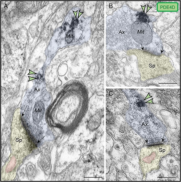
Presynaptic expression of PDE4D in ERC layer II. (A) PDE4D immunolabeling was observed in association with the plasma membrane in intervaricose axonal segments extending to form a glutamatergic-like axon terminal innervating a dendritic spine. (B-C) PDE4D is expressed in glutamatergic-like axonal terminals establishing axospinous Type I asymmetric synapses. The labeling is found in extrasynaptic subcompartments near the axon terminal plasmalemma, and not typically within the synaptic active zone. PDE4D immunolabeling in glutamatergic axon terminals was observed sometimes in close proximity to mitochondria (B). Synapses are between black arrows. Color-coded arrowheads point to PDE4D (green) immunoreactivity. Profiles are pseudocolored for clarity. Ax, axon; Sp, dendritic spine; Mit, mitochondria. Scale bars, 200 nm.