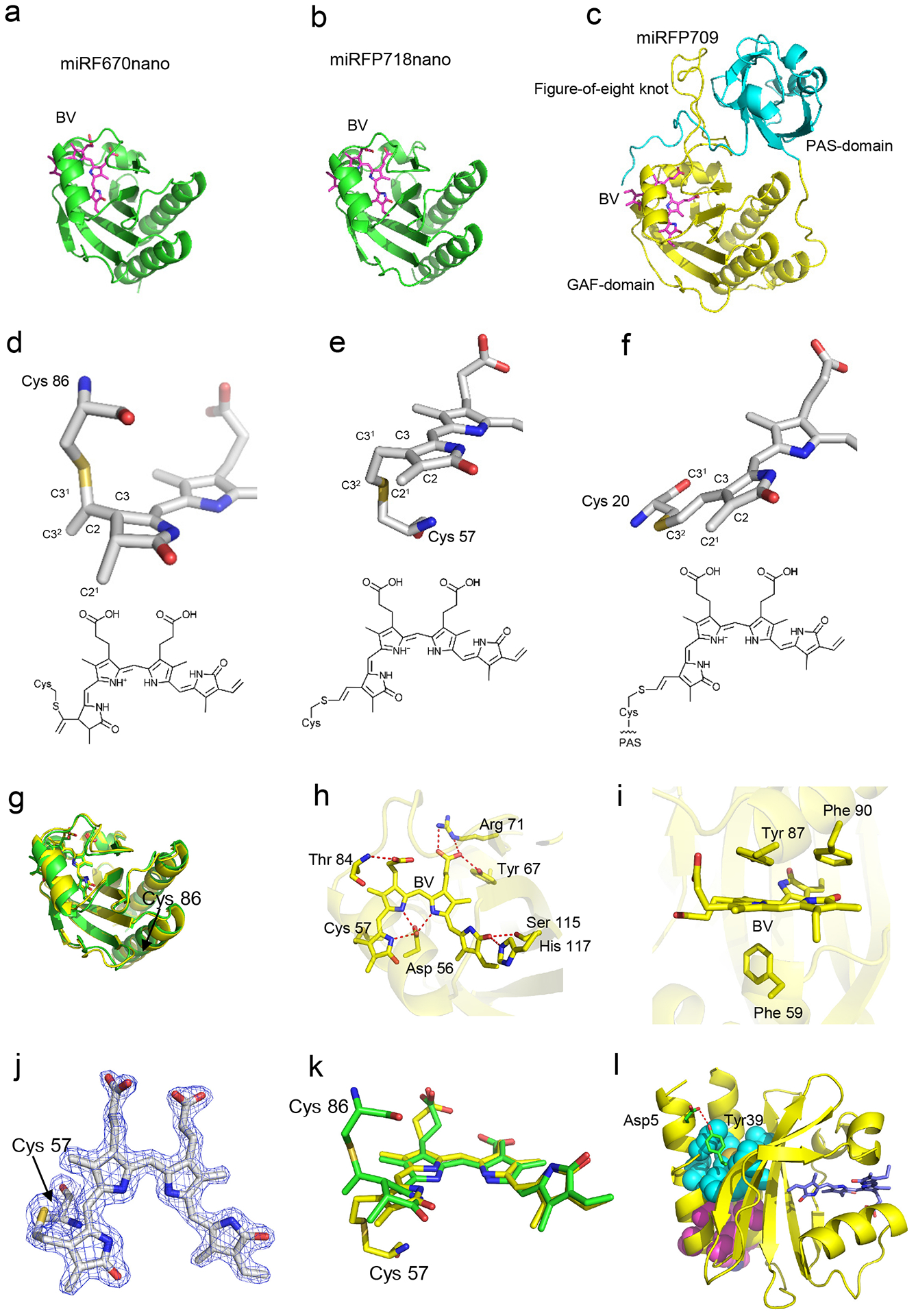
Extended Data Figure 1. Structure of miRFP718nano and its red-shifted chromophore.

Overall structures of (a) miRFP670nano (PDB ID: 6MGH), (b) miRFP718nano, and (c) miRFP709 (PDB ID: 5VIQ). The biliverdin (BV) chromophores are shown in magenta. The PAS and GAF domains of miRFP709 are in cyan and yellow, respectively. The BV chromophores in (d) miRFP670nano, (e) miRFP718nano, and (f) miRFP709 bound to the respective Cys residues and their chemical formulas. Carbon, nitrogen, oxygen, and sulfur atoms are in white, blue, red, and yellow, respectively. Sticks representations show only rings A and B of the chromophores and Cys residues. In miRFP670nano, the BV chromophore (d) is bound to Cys86 via the C31 atom, miRFP718nano (e) and miRFP709 (f) have the same chromophore species bound to the Cys57 and Cys20, respectively. (g) Superposition of miRFP670nano (green) and miRFP718nano (yellow) structures. (h) miRFP718nano hydrogen bond network around the chromophores (i) Stacking interactions between the chromophores and the surrounding residues in miRFP718nano. (j) The chromophores of miRFP718nano bound to the respective Cys57 residues in the 2Fo-Fc electron density map. The map is countered at 2.0σ-levels. (k) Superposition of the chromophores in miRFP670nano (green) and miRFP718nano (yellow). (l) Stabilizing mutations and hydrophobic clusters in miRFP718nano. The residues forming H-bonds are shown in green, hydrophobic clusters (one formed by residues Leu8, Ile11, Val12, Val26, Ile104, Leu114, Met140 and the other by Val15, Phe18, Leu19, Trp128, Phe132, Leu133) are in cyan and magenta.
