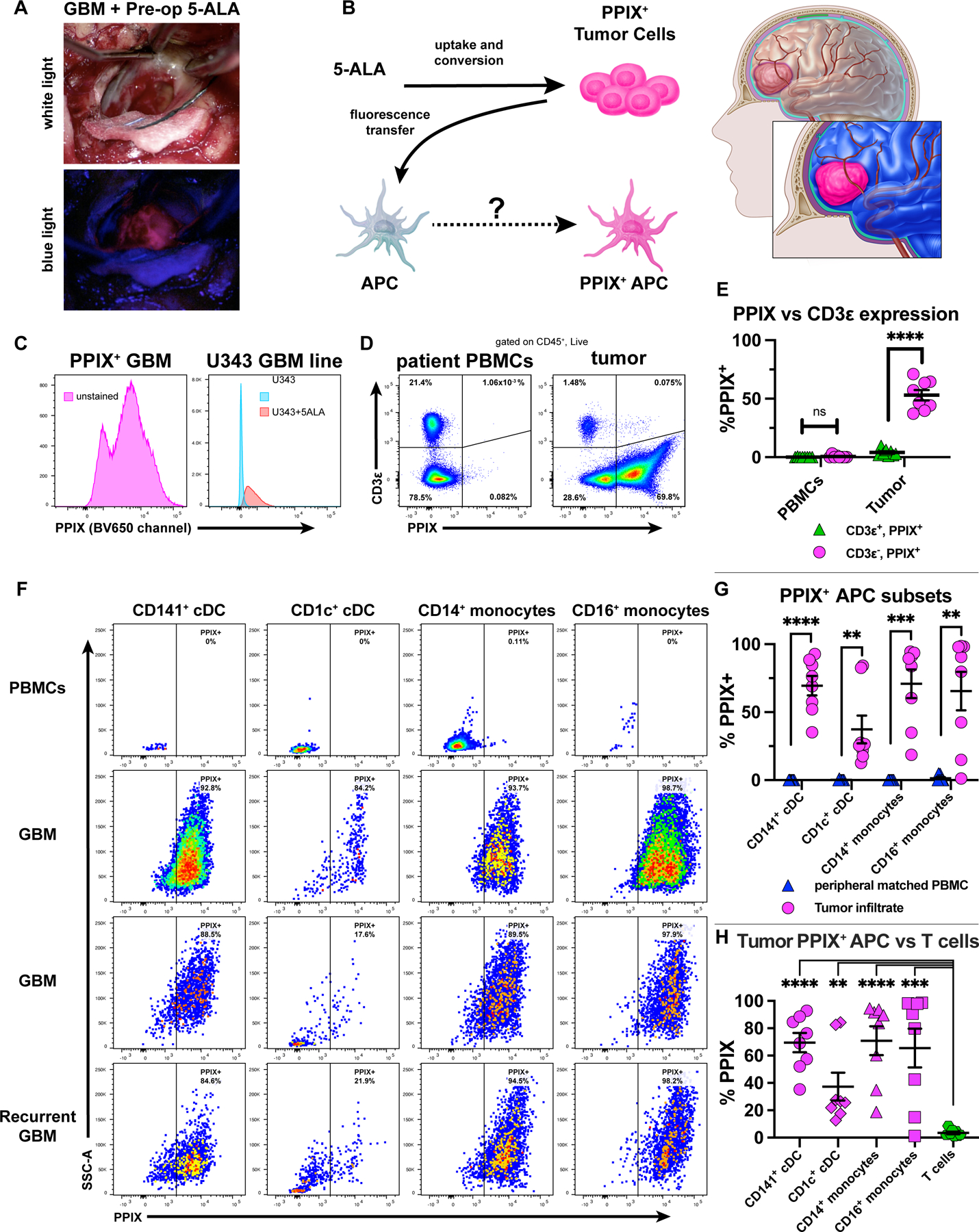
Figure 7. APCs infiltrating human GBM uptake the tumor-specific reporter PPIX.

(A) GBM patient with pre-administered 5-ALA: tumor visualized under white (top) or blue (bottom) light. (B) PPIX as tumor antigen surrogate, traceable in tumor-infiltrating APCs. (C) PPIX expression in a bulk unstained tumor from a 5-ALA-resected GBM (left) or in U343 cells treated with 5-ALA (right) (representative of independent experiments). (D-E) CD3ε vs. PPIX expression of live/CD45+ cells from patient-derived PBMCs or resected GBM tumors. (F-G) PPIX+ APC subsets across 3 GBM tumors (two primary, one recurrent) compared to patient PBMCs. (H) Tumor-infiltrating PPIX+ APCs vs. T-cells. Data representative of eight patients (six primary, two recurrent) in which GBM and matched intraoperative PBMCs were taken. Plotted are mean +/- SEM. **p<0.01, ***p<0.001, ****p<0.0001, ns: not significant. Unpaired two-tailed T tests for (E) and (H). Paired two-tailed T tests for (G).
