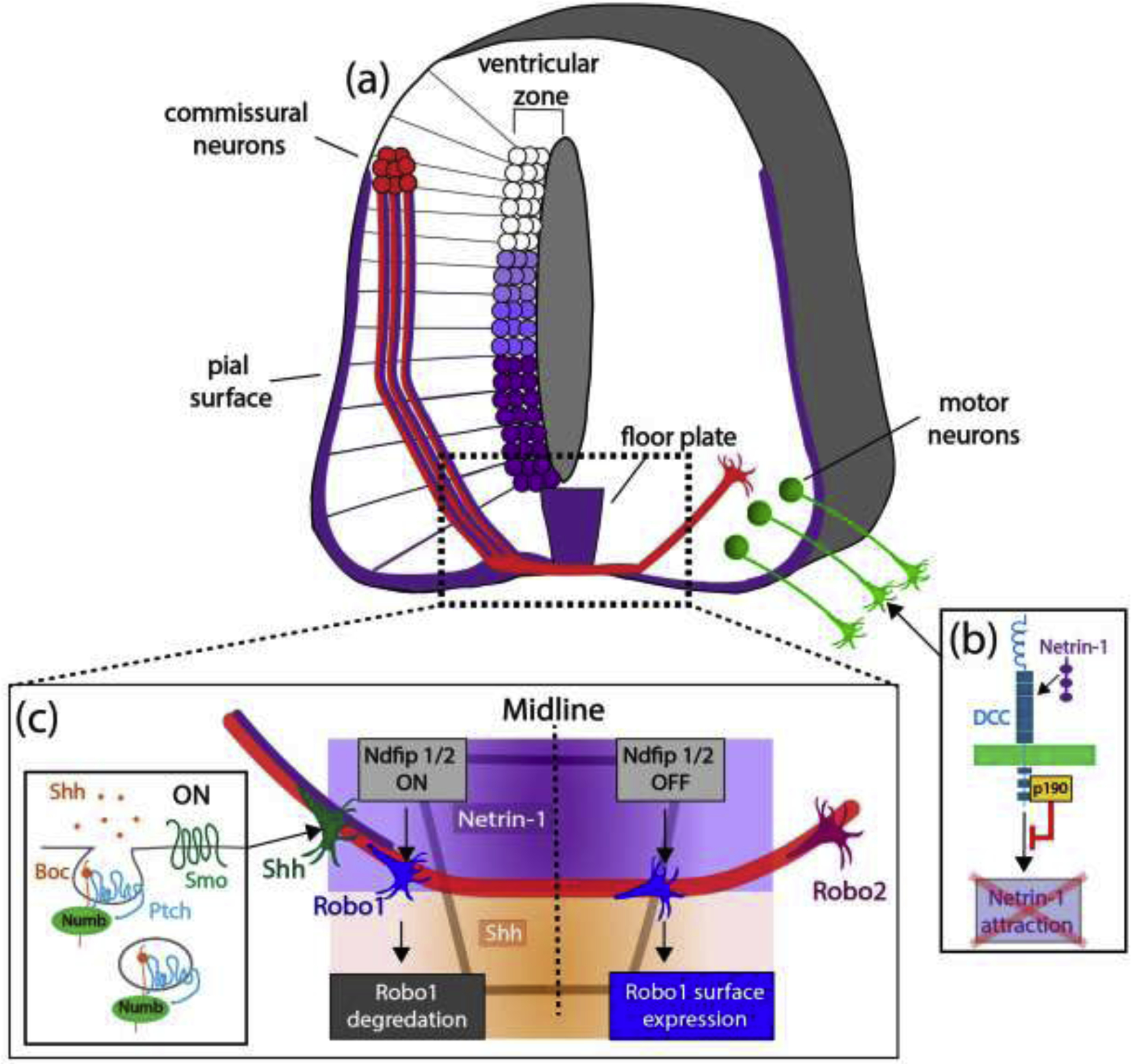
Figure 1. Molecular mechanisms underlying commissural and motor axon guidance in the developing spinal cord.

(a) Schematic of commissural axons navigating towards the floor plate (purple gradient: netrin-1 expression in the floorplate (FP), ventricular zone (VZ) and pial surface). Commissural neurons in the mouse dorsal spinal cord (red) extend their axons ventrally towards the floorplate (FP) and are attracted by netrin-1 (adapted from reference #6). (b) p190RhoGAP associates with DCC and prevents inappropriate attraction of spinal motor neuron axons to netrin-1, allowing exit from the spinal cord. (c) Schematic of molecular mechanisms underlying axon guidance at the midline. Shh secreted from the FP also attracts commissural axons (orange gradient). Shh-mediated endocytosis of the co-receptors Boc and Ptch1 (along with the adaptor Numb1) is necessary for Shh attraction. Robo1 (blue growth cones) and Robo2 (magenta growth cone) are differentially sorted to the cell surface and regulated during commissural guidance (see text for details).
