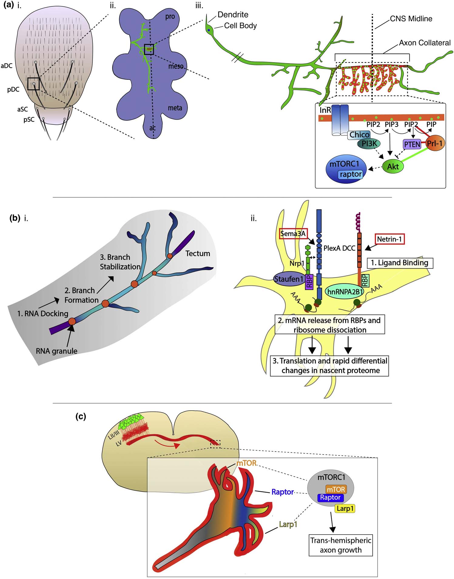
Figure 2. Subcellular regulation of axon guidance, branching and synapse formation.

(a) Subcellular regulation of single axon collateral branches and synapses by Prl-1 signaling. (i) Depiction of the Drosophila dorsal thorax with central sensory bristles: anterior (a)/posterior (p) dorsocentral (aDC/pDC) and scutellar (aSC/pSC). (ii) pDC axon (green) targeting the ventral nerve cord (VNC) (light purple). (iii) Enlarged view of the pDC neuron. Subcellular localization of Prl-1 regulates terminal arborization (via regulation of phosphoinositide signaling) (red) and synapse formation (green) specifically in the contralaterally-projecting axon collateral (green bars in the signaling diagram, synergistic genetic interaction; red bars, antagonistic genetic interaction). (adapted from reference #68) (b) Local translation regulates axon branching and guidance. (i) RNA granule (orange) docking precedes protrusion formation and stable axon branching in Xenopus RGC axons within the optics tectum. (ii) Specific guidance cue signaling can elicit rapid changes in the nascent proteasome of RGC axons. Nrp1 and DCC associate with distinct RNA-binding proteins (RBPs) that bind specific subsets of mRNAs. Upon ligand binding, ribosomes rapidly dissociate from their receptors, enabling mRNA release and subsequent translation, ultimately resulting in rapid and distinct changes to the nascent axonal proteome. (c) Growth cones at the tips of layer II/III callosal projection neuron axons in the mouse have transcriptomes and proteomes distinct from their somatic compartments. mTORC1 complex components are selectively enriched at the tips of trans-hemispheric growth cones, and this signaling is necessary for normal callosal projections.
