
Fig. 3.
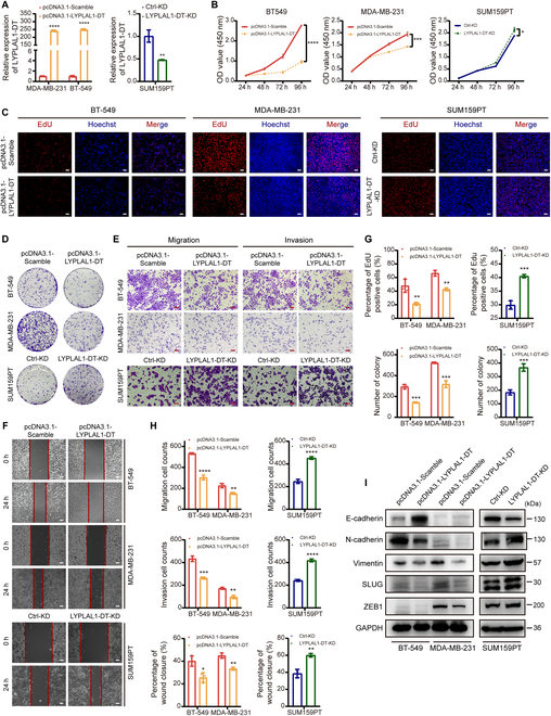
LYPLAL1-DT exerts antitumorigenic and antimetastatic effects to TNBC cells in vitro. (A) RT-qPCR result validating effects of overexpressing LYPLAL1-DT in MDA-MB-231 and BT-549 cells and knocking down LYPLAL1-DT in SUM159PT cells. (B) CCK-8 proliferation assays manifesting that overexpressing LYPLAL1-DT limited the proliferation of TNBC cells, while silencing LYPLAL1-DT promoted the proliferative nature. (C to F) Representative graphs of EdU staining proliferation assays (C), colony formation assays (D), transwell assays (E), and wound-healing assays (F) after performing overexpression or knockdown of LYPLAL1-DT in TNBC cells. (G) Quantitative data of EdU staining proliferation assays (top) and colony formation assays (bottom). (H) Quantitative data of transwell assays evaluating migration (top) or invasion (medium) properties and wound-healing assays (bottom). (I) Markers of epithelial-mesenchymal transition (EMT) were investigated in condition of overexpressing or knocking down LYPLAL1-DT in TNBC cells. Error bars represent mean ± SD. Scale bars in this figure all signify 100 μm. *P < 0.05, **P < 0.01, ***P < 0.001, ****P < 0.0001.