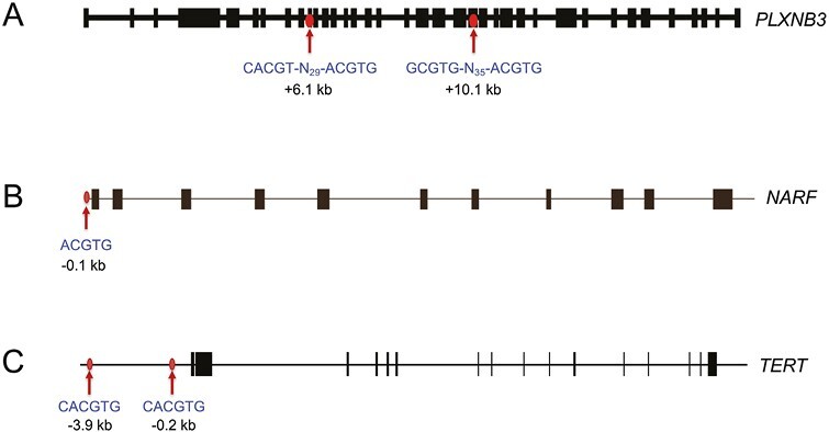
Figure 2.

Localization of HIF-1 binding sites in the PLXNB351 (A), NARF55 (B), and TERT56 (C) genes. Chromatin immunoprecipitation identified hypoxia-induced binding of HIF-1α and HIF-1β to genomic regions containing sequences (shown beneath the arrows) that match the HIF consensus binding site 5ʹ-RCGTG-3ʹ (R = A or G) or its complement 5ʹ-CACGY-3ʹ (Y = C or T). Note that these genes contain from 1 to 4 HIF consensus sequences in various orientations at locations (denoted by arrows) which are either upstream or downstream of the transcription start site (at the 5ʹ end of the first exon). Exon-intron structures of the genes are shown extending from left to right in the same orientation as the sequences.
