Figure 1.

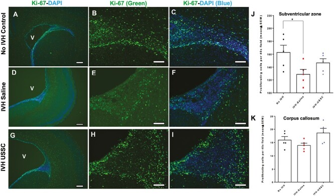
Impaired cell proliferation in the germinal matrix/corona radiata region of the subventricular zone (SVZ) after intraventricular hemorrhage at postnatal day 3. (A-I) Representative immunofluorescence images labeled with Ki-67 (proliferation marker, green) and counter stained with DAPI (nucleus marker, blue). Low magnification (4×) images from GM/CR (A, D, and G) and high magnification (20×) images with Ki-67 single stain (B, E, and H) and combined Ki-67 with DAPI (C, F, and I) in the 3 experimental groups indicated. Total proliferation immunofluorescence signal was lower in IVH + saline injected pups (middle panel, D-F) compared with no IVH healthy controls (upper panel, A-C) and a recovery in the USSC injected IVH premature pups (bottom panel, G-I). Sample size was 5 in each group, and 2-3 alternate coronal sections were taken at the level of mid-septal nucleus 20 µm sections. Scale bar = 100 µm. Ventricle (V) (J-K) Quantitative distribution of total proliferating cells in the subventricular zone (SVZ) and corpus callosum (CC). The scatter plot with bar graph showing mean cell density ± SEM among 3 experimental groups. Note: Significantly reduced proliferating cell density after IVH in SVZ but not in CC at day 3 (Control vs. IVH, P < .05). USSC treatment shows a trend to increase but not significant in both SVZ and CC; N = 5 in each group and cells were counted on 2-3 alternate coronal sections taken at the level of mid-septal nucleus. 20 µm sections.
