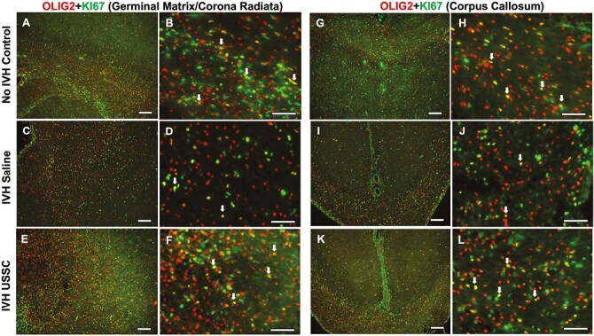
Figure 4.

USSC administration increased OLIG2 positive late progenitor cell proliferation in the GM/CR and CC after IVH on day 7. (A-F) Representative immunofluorescence images shown double-labeled coronal sections with Olig2 (red) and Ki-67 (green) in GM/CR. Low magnification images (10×) from healthy controls (A), IVH + saline (C) and USSC treated IVH pups (E). High magnification (40×) images double-labeled Ki67, Olig2 from controls (B), IVH + saline (D) and USSC treated IVH pups (F). Reduced double-positive immunosignals of OLIG2 with Ki-67 (yellow) in IVH (C, D) compared with both no IVH control (A, B) and IVH + USSC treated pups (E, F) in GM/CR of the SVZ. Double-positive cells shown with an arrow. Sample size of 5 in each group with 2-3 alternate coronal sections taken at the level of mid-septal nucleus of the forebrain for each pup; 20 µm sections. Scale bar = 100 µm. (G-L) Representative immunofluorescence images shown double labeled with Ki67-Olig2 in the corpus callosum. Low-magnification images (10×) from healthy controls (G), IVH saline (I), and USSC-treated IVH pups (K). High magnification (40×) images double-labeled Ki67-Olig2 from healthy controls (H), IVH + saline (J), and USSC-treated IVH pups (L). Reduced double-positive immunosignals of OLIG2 with Ki-67 (yellow) in IVH (I, J) compared with either no IVH control (G, H) or in IVH + USSC-treated pups (K, L) in CC region. Double-positive cells shown with an arrow. Sample size of 5 in each group with 2-3 alternate coronal sections taken at the level of mid-septal nucleus of the forebrain for each pup; 20µm sections. Scale bar = 100 µm.
