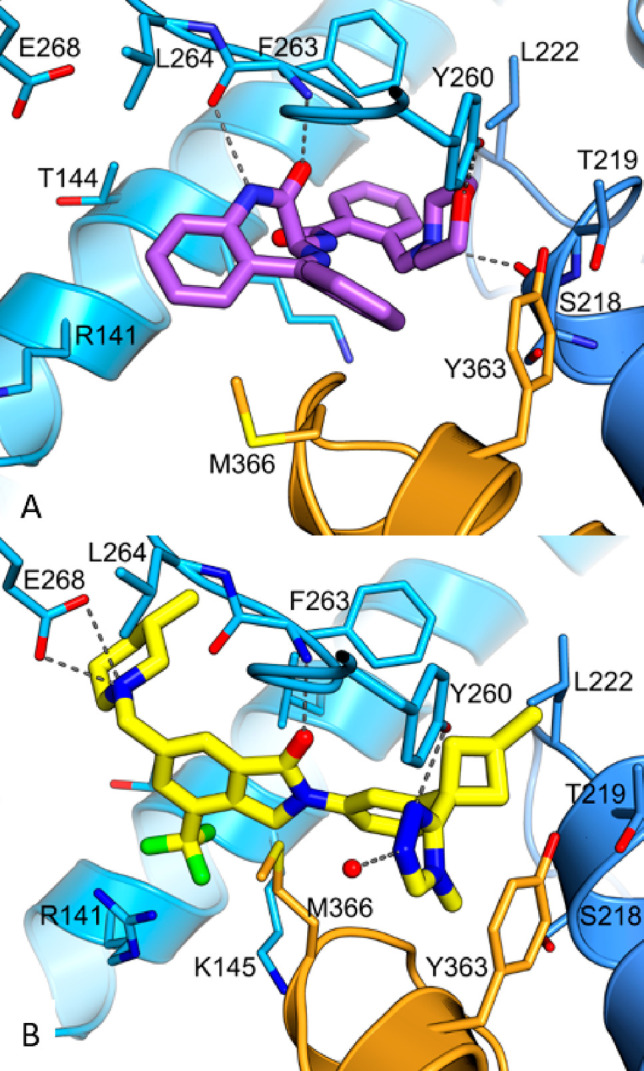
Figure 3.

(A) Crystal structure of compound 1 bound to the TKB/RING domain of Cbl-b (PDB code 8QNG). Compound 1 is colored purple, and the protein is in blue, with the linker helix region highlighted in orange. Hydrogen bonds are shown as dashed lines. (B) Crystal structure of Ex23 bound to the TKB/RING domain of Cbl-b (PDB code 8QNH). Ex23 is in yellow, and the protein is in blue, with the linker helix region highlighted in orange. Hydrogen bonds/ion-pair interactions are shown as dashed lines. Ex23 has recently been crystallized in another laboratory to show the same binding mode.34
