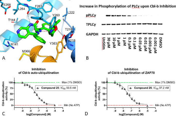
Figure 6.
(A) Crystal structure of compound 25 bound to the TKB/RING domain of Cbl-b (PDB code 8QNI). Compound 25 is in green, TKB domain in blue, and LHR in orange. Hydrogen bonds are shown as dashed lines; the putative intramolecular hydrogen bond is highlighted in red. (B) Western blot of a dose–response effect of Cbl-b inhibition on phosphorylation of PLCγ1 at Tyr783. Phosphorylated-PLCγ (pPLCγ) increases upon an increased dose of compound 25, in activated human T-cells. The left-most panel shows an unstimulated (UnStim) T-cell as a control, where the total PLCγ (TPLC) is completely unphosphorylated. (C) Inhibition of Cbl-b autoubiquitination via concentration-dependent dosing of compound 25. (D) Inhibition of Cbl-b ubiquitination of ZAP70 via concentration-dependent dosing of compound 25.

