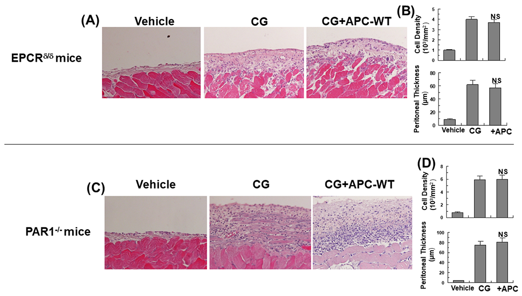
Figure 6. APC cannot inhibit CG-mediated PF in EPCR- and PAR1-deficient mice.

Representative light microscopic features of H&E staining of peritoneal tissues on day 21 in control (vehicle) mice, CG-injected mice treated with the vehicle alone, CG-injected mice treated with APC-WT in the EPCRδ/δ (A) and PAR1−/− (C) mice. Increased cell density and thickness of the submesothelial compact zone are presented for the CG-treated EPCRδ/δ (B) and PAR1−/− (D) mice. The data are shown as mean ± SEM, n= 10, *p<0.05 and **p<0.01. Scale bar =50μm.
