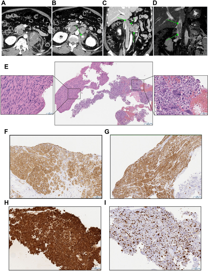
Fig. 2.

Radiological and histopathological characteristics in case 2. A-C An enhanced CT scan showing a 7 × 5 × 5 cm left-sided adrenal tumor with surrounding adipose tissue opacity, extravasation (A) and partial bleeding (B), as well as lymphadenopathy in the para-aortic region (C). D MRI revealed venous thrombosis extending from the left renal vein to the subhepatic vena cava. E Hematoxylin eosin staining showing atypical spindle cells and multinucleated cells in adrenal biopsies (40 x). F-H Tumor cell staining positive for smooth muscle actin SMA (F 100 x), desmin (G 100 x), and vimentin (H 100 x). I Immunohistochemical staining of the cells with the Ki-67 marker (100 x)
