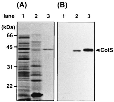
FIG. 1.
Detection of CotS protein by immunoblotting using anti-CotS antibody. The protein samples were solubilized from vegetative cells of B. subtilis 168trpC2 (lane 1), the SDS-mercaptoethanol-soluble fraction prepared from dormant spores of B. subtilis 168trpC2 (lane 2), and purified CotS protein with a His6 tag (lane 3). The samples were analyzed by SDS-PAGE (12% gel). (A) Coomassie brilliant blue stain; (B) immunoblotting using anti-CotS antibody. The arrowhead shows the migration position of CotS.

