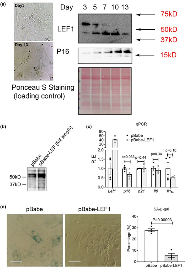
FIGURE 3.

LEF1 is implicated in cellular senescence. (a) Representative images of the morphology of MEFs at Day 3 and Day 13 after culturing. Immunoblot detection of p16 and LEF1 expression at different days. Ponceau S staining is provided as a loading control. (b) Immunoblot detection of LEF1 in cells expressing pBabe‐empty vector or pBabe‐LEF1 (long isoform) construct. (c) qPCR analysis of the expression of Lef1 and four common senescence markers at day 9 (8 days post retroviral infection) in MEFs. Results are shown as relative expression changes compared to cells transfected with the empty vector. (d) Representative images (left) and quantification (right) of SA‐β‐Gal staining in control or LEF1‐overexpressing MEFs at day 9 (8 days post retroviral infection).
