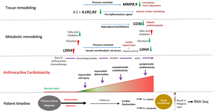
Figure 4. Schematic figure and timeline proposing the role of altered gene expression in anthracycline‐induced cardiomyopathy.

Progression of anthracycline‐induced cardiotoxicity involves a spectrum of remodeling at various levels, including structural, electrophysiological, metabolic, and functional events in the heart. Anthracycline exposure significantly reduces the expression of LDHA. Inhibition of LDHA obstructs aerobic glycolysis and activates fatty acid oxidation, which exacerbates cardiomyopathy under pressure overload. Hypoxia is a key regulator of cardiac hypertrophy and hypoxia‐inducible factor 1‐alpha activates transcription of LDHA, resulting in a second metabolic shift to aerobic glycolysis. Increase in CD36 facilitates the uptake of fatty acids and accumulation of lipids in cardiac muscle. Cardiac tissue remodeling involves activation of proinflammatory cytokines IL1R1 and IL1R2 that accelerate the progression of heart failure. MMP8 and MMP9 directly degrade extracellular matrix proteins, resulting in cardiomyocyte death and fibrosis. CD36 indicates cluster of differentiation 36; IL1R (1 and 2) interleukin 1 receptor type 1 and 2; LDHA, lactate dehydrogenase A; and MMP (8 and 9) matrix metalloproteinase 8 and 9.
