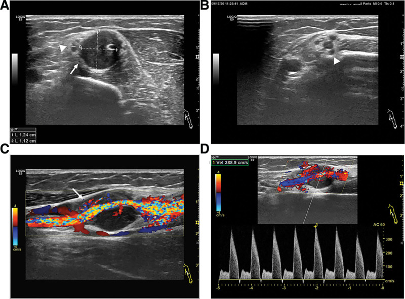
Figure 1.
Ultrasonography of the left brachial artery (BA). (A, B) A hypoechoic mass in the middle layer of the left BA (12 × 11mm), with regular morphology and clear boundaries (white arrow). The peripheral median nerve is compressed with edema (white arrowheads). (C) Longitudinal section examination reveals a hypoechoic mass between the outer wall and the lumen of the vessel with abundant blood flow running through, and the lumen is narrowed and distorted (white arrow). (D) Doppler ultrasound detects severe stenosis of the BA (the peak speed is 389cm/s).

