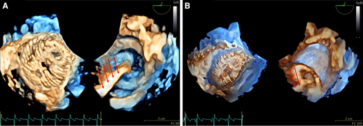
Figure 1.
3D real-time TOE of the atrial septal device before release in a 17 kg patient with a central atrial septal defect (ASD). (A) The figure demonstrates a 3D en face view of a 22 mm device from both the left atrium (left panel) and the right atrium (right panel) with the wire still attached (red arrows) (Supplementary Video S1). (B) Depiction of the “Minnesota maneuver” showing the pull on the right disk (Supplementary Video S2). 3D: three-dimensional; ASD, atrial septal defect; LA, left atrium; RA, right atrium; TOE, transoesophageal echocardiography.

