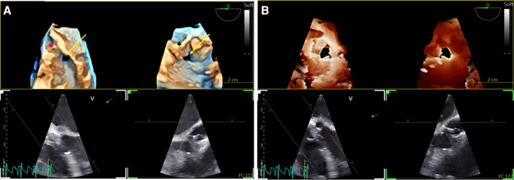
Figure 2.
3D TOE in a 5 kg patient with a ventricular septal defect (VSD). (A) Dual cropping views on 3D real-time of the perimembranous VSD with outlet extension (yellow arrow) from the LV close to the aortic valve (red asterix) (left panel) and from the RV close to the tricuspid valve (black asterix) (Supplementary Video S3). (B) Transillumination rendering in dual cropping views with Flexilight™ highlighting the perimembranous VSD with outlet extension (Supplementary Video S4). 3D, three-dimensional; LV, left ventricle; RV, right ventricle; VSD, ventricular septal defect.

