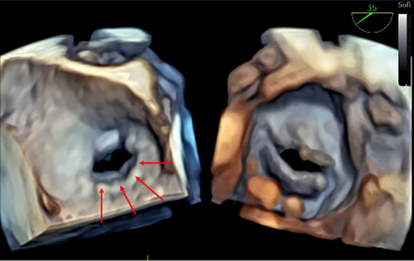
Figure 6.

3D TOE in a 37 kg patient with mitral stenosis. The figure displays dual cropping views on 3D real-time of the mitral valve. From the left atrium (left panel), a supravalvular ring is visible (red arrows), and from the left ventricle (right panel) (Supplementary Video S9). 3D, three-dimensional; LV, left ventricle.
