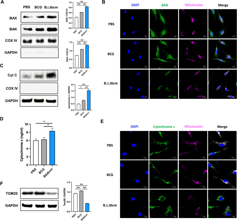
Fig. 5.
Deletion of tbcm exacerbates MOMP and cytochrome c release from mitochondria. BMDMs (BALB/c, female) were infected with BCG or BΔtbcm at an MOI of 10. A Expression levels of BAX and BAK were measured in the mitochondrial fraction of infected BMDMs at 6 h postinfection by western blotting. B Infected BMDMs were labeled with BAX and MitoTracker mitochondria 6 h postinfection. Confocal imaging of BAX translocation from the cytosol. C Cytosolic cytochrome c was detected by western blotting. D The release of cytochrome c was measured by ELISA in the cytosolic fraction at 8 h postinfection. E Infected BMDMs were stained with cytochrome c and MitoTracker at 8 h postinfection. Cytosolic cytochrome c was detected by confocal microscopy. E Western blotting of Tom20 expression levels in total cell lysates at 18 h postinfection. All data were collected in at least three independent experiments and are presented as the mean ± SEM. The results were analyzed for statistical significance by one-way ANOVA with Tukey’s multiple comparison test (*P < 0.05, **P < 0.01, ***P < 0.001). BCG, wild-type BCG; BΔtbcm, BCG Δtbcm mutant

