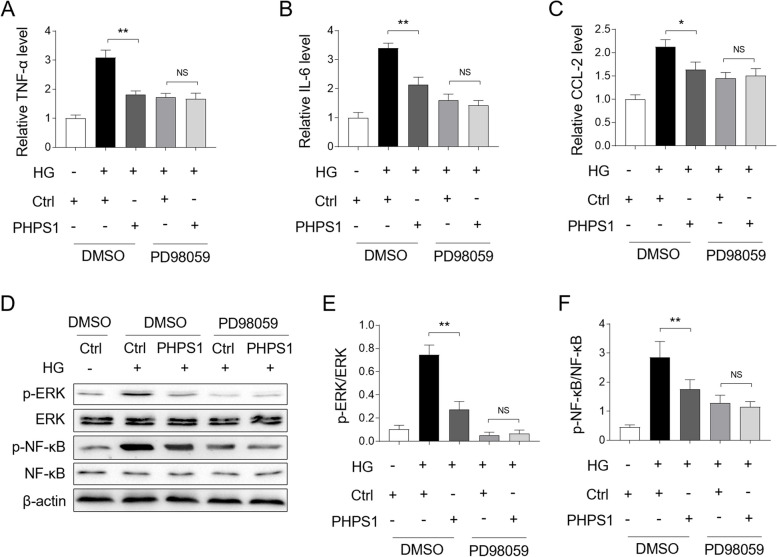
Fig. 6.
PHPS1 prevents high glucose-induced inflammatory responses through ERK/NF-κB pathway. A-C HK-2 cells were pretreated with 5 μM PHPS1 alone or in combination with 50 μM PD98059 before exposure to high glucose (HG, 30 mM) for 24 h. The control group was incubated with normal glucose (NG, 5.5 mM). The mRNA level of TNF-α (A), IL-6 (B), and CCL-2 (C) in each group of HK-2 cells was quantified by qRT-PCR analysis. Student’s test, **, p < 0.01; *, p < 0.05; NS, not significant (n = 3). D-F HK-2 cells were treated as in (A-C), the levels of p-ERK, p-NF-κB and their basal protein expression were measured by Western blot analysis. The representative images were shown (D). The intensity of potein bands (D) was quntified by ImageJ and the ratio of p-ERK/ERK (E) and p-NF-κB/ NF-κB (F) were depicted. Student’s test, **, p < 0.01; NS, not significant (n = 3)

