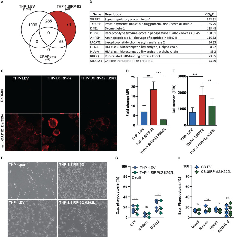
Figure 4.
SIRP-ß2 with a point mutation in the charged lysine residue at position 202 (SIRP-ß2.K202L) in THP-1 cells did not augment adhesive and phagocytic effects. (A) Schematic GFP immunoprecipitation (IP), of THP-1.EV vs. THP-1.SIRP-ß2, including 127 unique protein hits of THP1-SIRP-ß2. (B) List of the 10 most frequently occurring proteins found in THP-1.SIRP-ß2, DAP12 as a leading hit. (C) Representative confocal microscopy images of anti-DAP12 staining on THP-1.EV, THP-1.SIRP-ß2 and THP-1.SIRP-ß2.K202L. (D) DAP12 expression on THP-1.EV, THP-1.SIRP-ß2 and THP-1.SIRP-ß2.K202L, determined by flow cytometry (n=3). (E) Quantitative adherent cells in field of view (FOV) of THP-1.EV, THP-1.SIRP-ß2 and THP-1.SIRP-ß2.K202L. (F) Representative images of adherent THP-1.parental, THP-1.EV, THP-1.SIRP-ß2 and THP-1.SIRP-ß2.K202L. (G) Experimental THP-1 mediated phagocytosis with Daudi cells for 3h, including RTX, Inhibrx and B6H12 treatment (1 µg/ml) (n=3). (H) Experimental CB-derived macrophage phagocytosis of EV vs. SIRP-ß2.K202L with Daudi, Ramos, U2932, SUDHL-6. p values are indicated as: *** p < 0.001 and ** p < 0.01. ns (not significant).

