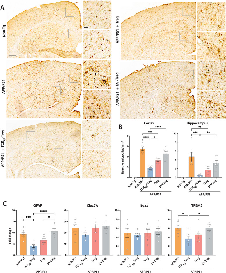
Fig. 6.
Adoptive transfer of TCRAβ-Tregs reduces reactive microglia in APP/PS1 mice. A APP/PS1 mice were untreated or treated with TCRAβ-Tregs, polyclonal Tregs (Treg) or EV-Tregs (TCR−−-Tregs electroporated with empty plasmid vector) by adoptive transfer and brain tissues acquired 3 weeks post-transfer. Untreated non-transgenic mice served as controls. Representative images showing Iba1 reactive cells in brain regions. Scale bar = 100 µm. Areas with most Iba1+ reactive microglia are highlighted by inserts for the cortex and hippocampus. Scale bar = 50 µm. B Number of Iba1+ reactive microglia were quantified from immunohistochemistry in cortex and hippocampus using Optical Fractionator probe of Stereo Investigator. Data presented as mean ± SEM for 5–6 mice per group. C Changes in the expression of the disease-associated genes for astrocytes (GFAP) and microglia (Clec7A, Itgax, and TREM2) in cortical tissue by qPCR. Obtained CT values were normalized against the RPLP0 gene and non-Tg mice was used as control. Data presented as mean ± SEM. One-way ANOVA followed by Turkey’s post hoc test was used to determine significant differences between experimental groups. *p < 0.05, **p < 0.01, ***p < 0.001, ****p < 0.0001

