Fig. 3.
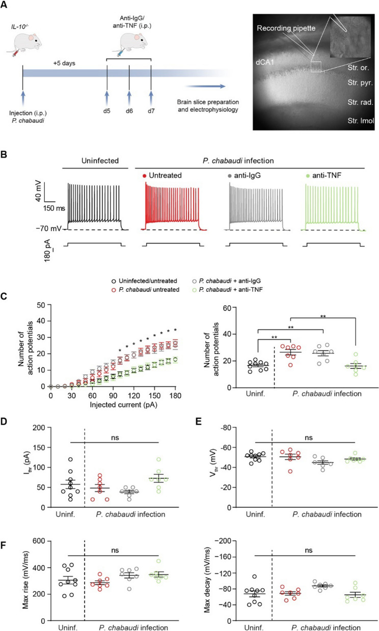
In vivo treatment with anti-TNF antibody mitigates neuronal hyperexcitability in eCM. A Left: timeline for infection, antibody treatment, and electrophysiological recordings. Right: representation of patch-clamp recording of CA1 pyramidal neuron. B Representative traces of action potentials fired by CA1 neurons in slices from the indicated experimental group at the 180-pA injected current step. C Number of action potentials fired by CA1 neurons over a range of injected current stimuli. D, E Comparison of the current (D) and voltage (E) thresholds for action potential initiation among the indicated experimental groups. F Comparison of action potential kinetics among the indicated experimental groups. Data are mean ± SEM (n = 7–9 neurons/group; slices from at least N = 3 mice per group). Significance was assessed using a one-way ANOVA with post hoc Tukey’s multiple comparisons test. In (C, right, D, E, and F): ns, not significant; **p < 0.01. In (C, left), * denotes current steps at which the number of action potentials fired by CA1 neurons of infected mice and infected mice treated with the isotype-matched control antibody is significantly greater (p is at least < 0.05) than the number of action potentials fired by CA1 neurons of uninfected mice and infected mice treated with the anti-TNF antibody

