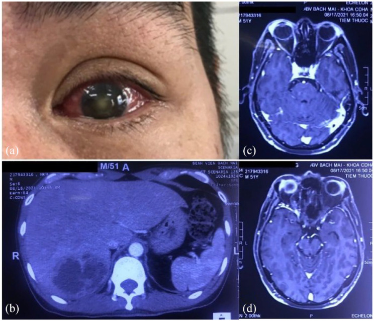
Figure 1.
Eye involvement and pyogenic liver abscess of patient one. (a) OS involvement. (b) Contrast-enhanced abdominal computerized tomography (CT) scanning showed hepatomegaly with 19.5 cm. The posterior parenchymal segment adjacent to the diaphragm has heterogeneous hypoechoic foci of size 70 × 55 × 50 mm, cortical enhancement, central liquefaction, and perfusion disturbances in the liver parenchyma around the lesion. (c and d) On magnetic resonance imaging, there is a component of increased signal on T1W, decreased T2W, limited diffusion, and no enhancement after injection. Infiltrate the orbital soft tissue around the eyeball and the lateral rectus muscle on OS, enhancing after injection.

