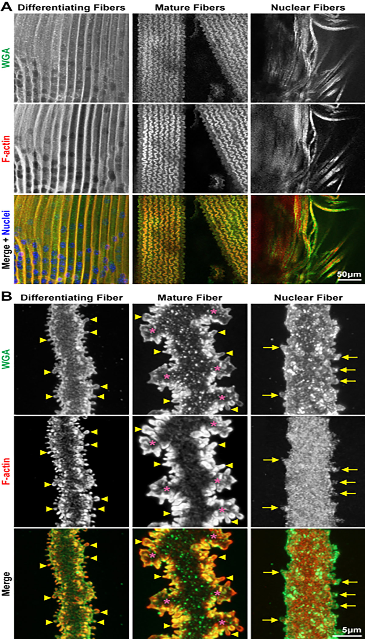
Figure 3: Confocal images of bundles (low-magnification) and single (high-magnification) images of fiber cells from different regions of the lens.

Cells are stained with WGA (cell membranes, green), phalloidin (F-actin, red), and DAPI (nuclei, blue). (A) In these preparations, bundles of lens fiber cells are often found from different regions. In the cortical preparations, there are differentiating and mature fiber cells. These different cells have distinct morphologies, allowing for easy identification. In the samples from the lens nucleus, fiber cell bundles as well as dissociated fibers are found. Scale bar = 50 μm. (B) Z-stacks through single differentiating, mature, and nuclear fiber cells are collected, and 3D rendering is used to reconstruct the images shown. The differentiating and mature fibers have many small protrusions along their short sides (the arrowheads mark select ones). These small protrusions are enriched for F-actin. The mature fiber also has large interlocking paddle domains (asterisks). The nuclear fibers have small membrane pockets and divots stained by WGA and retain a few protrusions along their short sides (arrows). Surprisingly, the F-actin network is now distributed in the cell cytoplasm without enriched signals at the cell membrane. The F-actin network does extend into small protrusions. Scale bar = 5 μm.
