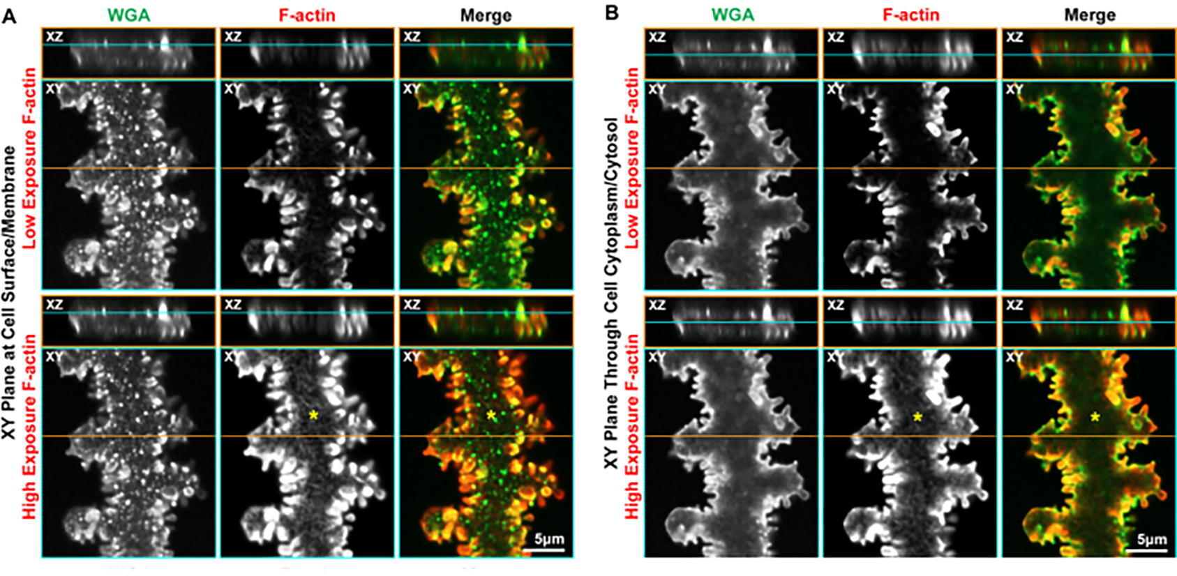
Figure 5: Orthogonal 2D projections (XY and XZ planes) of 3D confocal z-stacks through a single mature fiber cell stained with WGA (green) and phalloidin (F-actin, red).

(A) Projections from the cell surface along the broad side show WGA along the cell membrane, and the puncta of WGA suggest the membrane is not completely smooth. F-actin is enriched in the protrusions along the short sides, and a high-exposure image of the F-actin staining reveals a reticulated network along the cell membrane (asterisks). (B) Another plane through the cytoplasm of the same cell shows WGA along the cell membrane, framing the cell. F-actin forms a reticulated network through the cytoplasm (asterisks) and is greatly enriched in the protrusions along the cell membrane. Scale bars = 5 μm.
