Fig. 5.
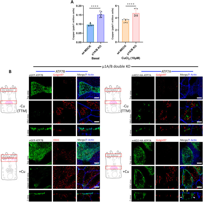
AP-1 is crucial for TGN retention of ATP7A and ATP7B. (A) Intracellular copper level in wild-type (wt) and AP-1 KO MDCK cells under basal and copper-treated conditions. AP-1 KO cells show higher copper levels in both conditions [i.e. basal and copper treated cells (10 µM CuCl2 for 4 h)] compared to wild-type MDCK cells. Results are mean±s.d (n=3). ****P<0.0001 (Mann–Whitney U-test/Wilcoxon rank-sum test). (B) Polarized AP-1 KO MDCK cells (µ1A and µ1B double-knockout cells) showing localization of transfected mKO2–HA–ATP7A and eGFP–ATP7B. ATP7B has lost its TGN retention and constitutively localizes to the apical surface, retaining its polarity in copper-depleted as well as copper-treated conditions. ATP7A has also lost its TGN localization and was found to be vesicularized in a copper-chelated environment, but in copper-treated medium it localizes to both the apical (yellow arrowhead) as well as the basolateral surface (white arrowhead), losing its polarity. Images representative of five repeats. Scale bars: 5 µm.

