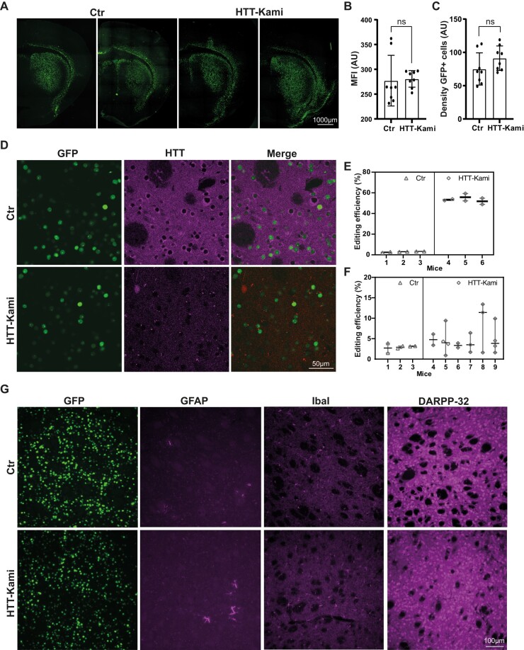
Figure 5.
Normal brain features following HTT inactivation in adult FVB mice. (A) Two representative striatal sections from 9.5-month-old Ctr and Htt-KamiCas9 mice showing nuclear expression of the GFP encoded by the AAV vectors. (B and C) Quantitative analysis confirmed that the mean fluorescence intensity (MFI in arbitrary unit: AU) (B) and the density of GFP-positive cells (C) were similar in the two groups (n = 4 animals/group). Statistics: two-tailed t-test (MFI: P = 0.8539, t(0.1876), df(14), density: P = 0.1528, t(1.512), df(14)). (D) Confocal images showing the loss of Htt in GFP-positive striatal cells. Cortical (n = 3 animals/Ctr, n = 6/treated animals; 2–4 punches/animal) (E) and striatal Htt editing (n = 3 mice/group, 2 punches/animal) (F) in 11-month-old mice. Statistics: nested two-tailed t-test (cortex: P = 0.1658, t(1.433), df(22), striatum: P < 0.0001, t(35,5), df(10)). (G) Immunostaining for glial fibrillary acidic protein (GFAP), ionized calcium binding adaptor molecule 1 (IbaI) and dopamine- and cyclic-AMP-regulated phosphoprotein of molecular weight 32 000 (DARPP-32) in Ctr and Htt-kamiCas9 mice.

