
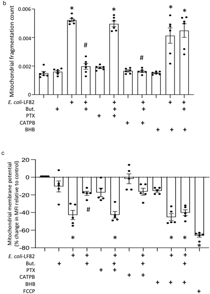
Figure 7.
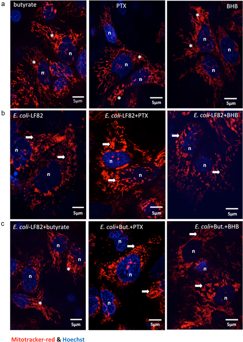
Pertussis toxin and -hydroxybutyrate (BHB) implicate FFAR3 in butyrate’s maintenance of epithelial mitochondrial networks. Monolayers of the human colon-derived T84 epithelial cell line (106) were treated with E. coli-LF82 (108 cfu, 4 h) ± a co-treatment with sodium butyrate (But., 10 mM) ± an 18 h pre-treatment with pertussis toxin (PTX, 50 ng/ml) or BHB (5 mM). (a) Representative images were collected in a random fashion by first identifying epithelia nuclei (blue, n) and then swapping the confocal laser channel to assess the mitochondrial network (Mitotracker (red)). Twenty cells per monolayer were characterized for mitochondrial fragmentation count by ImageJ analysis and averaged/monolayer (b). (c) Mitochondrial membrane potential was assessed by TMRE fluorescence in a flow cytometer. A 10 min treatment with the metabolic toxin, FCCP (10 Ohms.cm2) was used as a positive control. The effect of the FFAR2 antagonist (S)-3-(2-(3-chlorophenyl) acetamido)-4-(4-(trifluoromethyl) phenyl) butanoic acid (CATPB, 10 Ohms.cm2, 30 min pre-treatment) is also shown (mean ± SEM; n = 5–6 epithelial monolayers from separate experiments in panels a and b; * and #, p <.05 compared to control uninfected cells and E. coli-LF82 only infected cells respectively by two-way ANOVA followed by Tukey’s multiple comparison test (b) or the Kruskal–Wallis test from by Dunn’s multiple comparison test; image *, fused mitochondrial network with elongated strands; arrow, fragmented, vesiculated area of the mitochondrial network; MFI, mean fluorescence intensity).
Figure 7.
(continue).