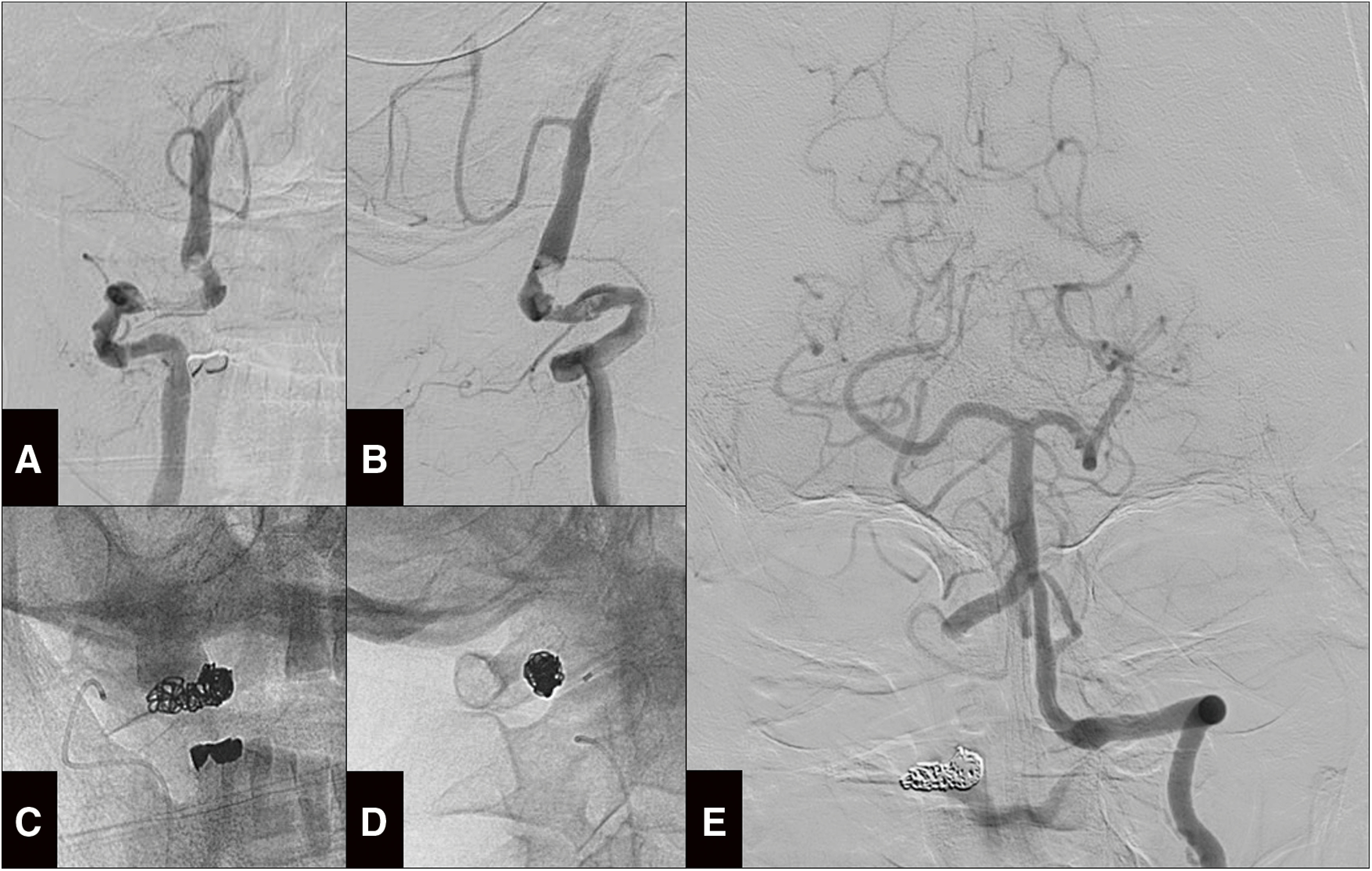
Fig. 4. PAO for the right dissected VA on the fifth day of hospitalization. (A and B) Preoperative right vertebral angiography from the anteroposterior (A) and lateral (B) views showing the stagnating flow and persistent V3 dissection. (C and D) The coil mass placed on the V3 segment on the anteroposterior (C) and lateral (D) views. (E) Postoperative left vertebral angiography demonstrating normal flow to the right posterior inferior cerebellar artery.

