FIG. 1.
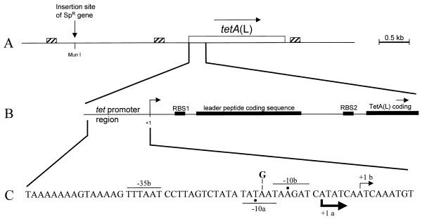
Map of the tetA(L) chromosomal locus. (A) Schematic diagram of the 4.5-kb HindIII fragment cloned into pJBC1 (4). The location of the MunI site, into which the Spr gene was inserted, is shown. Small boxes with diagonal stripes mark the locations of homologous 170-bp direct repeats (1). Open box, tetA(L) gene. (B) Expanded view of the tetA(L) promoter and leader region. Filled boxes, translational signals and coding sequences. The site of transcription initiation is marked +1. RBS, ribosome binding site. (C) Nucleotide sequence of the tetA(L) promoter region. Transcriptional start sites (+1) a and b are indicated, along with the respective −10 and −35 sequences. Actual −10 positions are marked by dots. The thicknesses of the arrows showing the direction of transcription indicate the relative amounts of transcription initiation from the two start sites in the wild type. The location of the A→G change at position −14 (relative to start site b) is shown.

