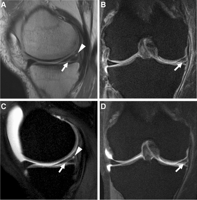
Fig. 21.
Recurrent meniscal tear. Fifty-one-year-old man with history of meniscal surgery and recurrent symptoms. Sagittal intermediate-weighted (A) and coronal intermediate-weighted fat-suppressed (B) conventional MR images show evidence of partial medial meniscectomy with linear increased signal extending to the inferior surface (arrows) and small posterior parameniscal cyst (arrowhead), consistent with a recurrent tear. Sagittal (C) and coronal (D) T1-weighted fat-suppressed MR arthrogram images demonstrate dilute gadolinium extending through the tear (arrows) and filling the parameniscal cyst (arrowhead)

