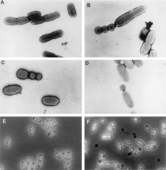
FIG. 1.
(A to D) Electron micrographs. Cells were fixed on an electron microscopy grid with 1% phosphotungstic acid and observed at a magnification of ×5,000. Strains: JP406 (cpxA+), A; JP408 (cpxA9*), B to D. (E and F) Phase-contrast micrographs. Division products without DNA appear as dark bodies. Strains: JP406 (cpxA+), E; JP467 (cpxA2.1*), F.

