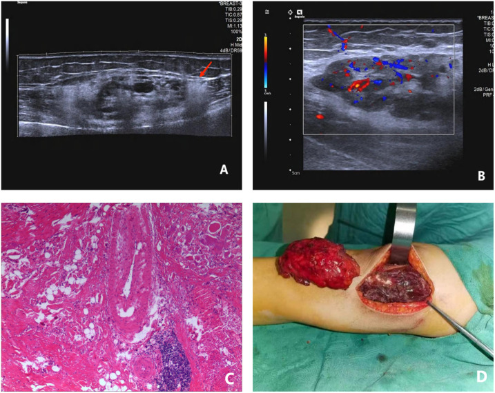
Figure 4.
A female patient, 12 years old and suffering from intermuscular FAVA in the right thigh; (A) longitudinal 2D ultrasound image showed a mixed intermuscular solid mass with triangular hyperechoic “fascialtail sign” (red arrow) at the lower edge of the lesion; (B) through enlargement of local view of the lesion, CDFI suggested a small amount of blood flow signal within the lesion; (C) pathological findings showed existence of abnormally dilated vessels, fatty tissues and fibrous tissues in muscle cells. Fiber content could be found around the lesion, HE×100; (D) a bright red tumor with a tough texture was found during the operation in the deep layer of the right thigh muscle. FAVA, fibro adipose vascular anomaly.

