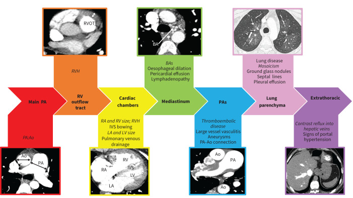
FIGURE 1.
Systematic approach to computed tomography pulmonary angiogram interpretation. Ao: aorta; BA: bronchial artery; IVS: interventricular septum; LA: left atrium; LV: left ventricle; PA: pulmonary artery; RA: right atrium; RV: right ventricle; RVH: right ventricular hypertrophy; RVOT: right ventricular outflow tract. Main features demonstrated in each image are in italics.

