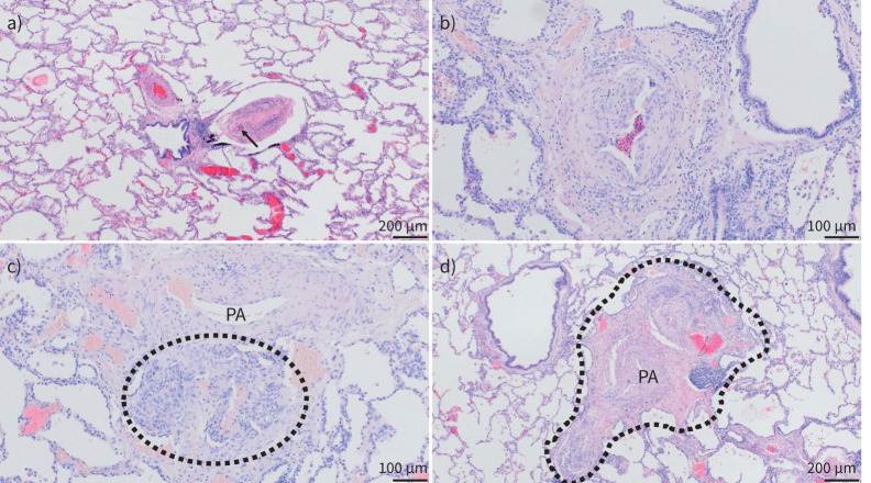
FIGURE 2.
Pulmonary vascular histopathology in idiopathic and hereditary pulmonary arterial hypertension (PAH). Lungs from patients suffering from idiopathic and hereditary PAH; haematoxylin and eosin staining. a) Well-preserved lung parenchyma with slender alveolar septa and extended alveolar spaces; note the central bronchiole with two pulmonary arterial (PA) branches: the left one with medial and intimal concentric thickening, the right one displaying near-occlusive concentric intimal fibrosis (arrow indicates remaining lumen) and moderate lymphocytic infiltrate. b) PA with eccentric intimal fibrosis and cushion-like intimal thickening, suggesting an organised thrombotic event or in situ thrombosis. c) Plexiform lesion (circled) arising from the adventitia of a PA (top) and representing a connection between systemic vasa vasorum and pulmonary vasculature. d) Singular millimetric fibro-vascular lesion in a patient with hereditary PAH (bone morphogenetic protein type II receptor mutation); this vascular and fibrotic conglomerate comprising systemic and pulmonary vessels measures about 1.5 mm (circled area). a) and d) magnification ×40; b) and c) magnification ×100.

