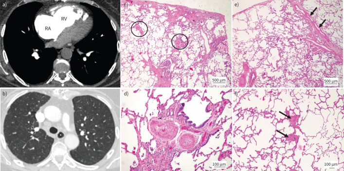
FIGURE 4.
Pulmonary arterial hypertension (PAH) in association with systemic sclerosis. a) Dilated right atrium (RA) and right ventricle (RV). b) Prominent oesophagus (*). c–f) Lungs from a different patient suffering from systemic sclerosis-associated PAH. Haematoxylin and eosin staining; magnification: c) and e) ×20; d) ×100; and f) ×40. c) Peripheral lung parenchyma with mild subpleural fibrosis (top). Note heavily remodelled muscular-type pulmonary arteries (PAs) (circled) in preserved areas, independent of fibrosis. d) Magnification of right circled PA. Medial muscular thickening and important near-occlusive concentric nonlaminar intimal fibrosis is present. e) Remodelled PAs (bottom left) and septal veins with substantial smooth muscle cell hyperplasia (arrows). f) Muscularisation of small pulmonary arterioles (arrows) and thickening of alveolar septa to the left of the arterioles; these changes are vaguely reminiscent of PAH group 1.6 (pulmonary veno-occlusive disease/pulmonary capillary haemangiomatosis).

