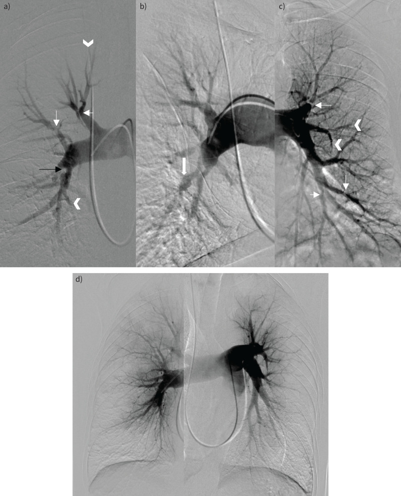
FIGURE 4.
Digital subtraction angiography. a) Patient 1. Pulmonary vascular signs of chronic thromboembolic disease. There are bilateral segmental webs and stenoses (thin white arrows in panels a and c), post-stenotic dilatation (block arrow in b), intimal irregularity (black arrow in a), multifocal occlusions, and abrupt vessel truncation (arrowheads in a and c). d) Patient 2. Diffuse narrowing of the subsegmental vessels with abrupt calibre change. The distal vessels are spindly with a corresponding peripheral perfusional defect. There was no significant disease in the proximal vessels.

