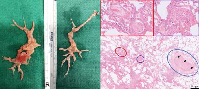
FIGURE 6.
Left panel: resected material during pulmonary endarterectomy in patient 1. Right panel: lungs from another patient with chronic thromboembolic pulmonary hypertension with peripheral, inoperable disease who underwent a lung transplantation (haematoxylin and eosin staining; magnification: ×40, insets ×200). A pulmonary artery of a cross-sectional diameter of about 400 µm (circled in blue) is obliterated by organised thromboembolic material. Note the recanalisation of vessels within the obstruction (arrows). In the vicinity of this obstructed artery, microvascular remodelling can be found in arterioles (circled in purple), as well as in post-capillary venules (circled in red), both of around 70 µm in diameter, and depicted in the magnified purple and red inset boxes above. Note the concentric character of the microvascular remodelling, which indicates reactive changes, rather than thrombotic lesion, with smooth muscle cell hyperplasia and intimal fibrosis (venules).

