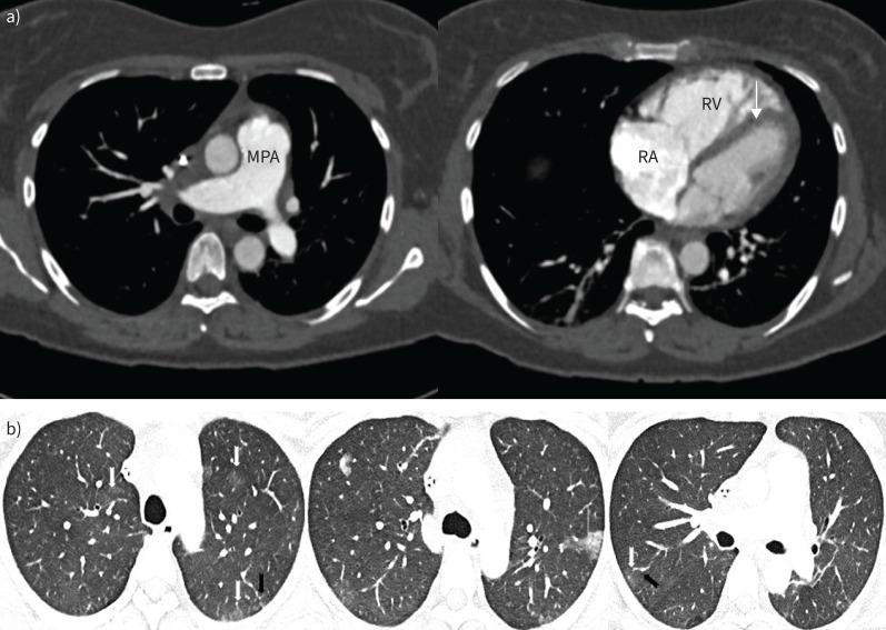
FIGURE 8.
a) Axial computed tomography (CT) images showing dilated main pulmonary artery (MPA), mildly dilated right atrium (RA) and mildly dilated right ventricle (RV) with flattening of the interventricular septum (arrow). b) Lung windows from the same CT demonstrating widespread lung parenchymal abnormalities. There are multifocal subtle ground glass opacities (white arrows), a few dilated subsegmental arteries (black arrows) and subpleural consolidation (middle panel). b) Reproduced from [25].

