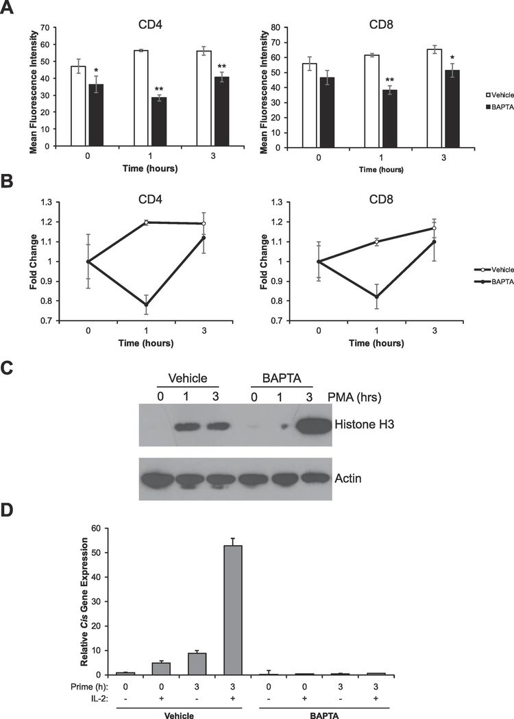
Figure 3: A calcium-dependent PKC is required for proper chromatin decondensation and acquisition of competence to respond to IL-2.
(A) Splenocytes were treated with vehicle (DMSO) or 10μM BAPTA-AM for 30 minutes. Cells were then stimulated with 10ng/mL PMA for the times indicated. Cells were then analyzed by flow cytometry to determine chromatin accessibility in CD4+ and CD8+ cells by intracellular staining for H3K4me1. Data are the means ± SD of triplicates and are presented as the mean fluorescence intensity of H3K4me1 staining. *p < 0.05, **p < 0.001 (Student’s t-test) compared with the vehicle control at each time point. (B) Data from A was calibrated to the 0 hour time point to show fold change in chromatin decondensation over time. (C) Sorted naïve (CD25-) T-cells were left untreated or stimulated with vehicle (DMSO) or 10μM BAPTA-AM for 30 minutes. Cells were then stimulated with 10ng/mL PMA for the times indicated. Proteins were resolved via SDS-PAGE and histone solubility was measured via detection of histone H3 as assayed by Western blot. Detection of Actin serves as a loading control. (D) Sorted naive (CD25-) T cells were left untreated (naïve) or treated with vehicle (DMSO) or 10μM BAPTA-AM for 30 minutes, then cells were stimulated with 10ng/mL PMA for the times indicated. Each sample was then divided in half and either left untreated or stimulated with 1000U/mL of IL-2 for 1 hour. Total RNA was isolated and expression of the STAT5 target gene Cis was determined by qRT-PCR relative to the housekeeping gene CD3ε. Data were calibrated to the naïve 0 hour control. All data are representative of at least 3 independent experiments.

