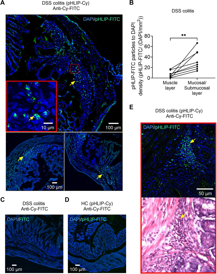
Fig. 3: pHLIP sublocalization in the colon of healthy control and acute DSS colitis mice.

A. DAPI/ anti-Cy-FITC labelling in DSS colitis mice treated with pHLIP (representative images of N= 2 mice). Staining (arrows) is predominantly localized in the mucosal and submucosal layer. Inset shows the subcellular localization of pHLIP. See also Supplementary digital content Fig. 2 for expanded confocal images illustrating the insertion of pHLIP into cell membranes within the inflamed layers of the colon. B. pHLIP-FITC particle distribution within the colon wall of DSS colitis mice (N=8). C. DAPI/ anti-Cy-FITC labelling in a DSS colitis mouse that was not treated with pHLIP. D. DAPI/ anti-Cy-FITC labelling in a healthy control (HC) mouse treated with pHLIP. E. DAPI/ anti-Cy-FITC labelling and H&E staining in a DSS colitis mouse treated with pHLIP. Arrows denote the co-localization of pHLIP particles and infiltrating inflammatory cells. Abbreviations: Cy, Cyanin. DAPI, 4′,6-diamidino-2-phenylindole. DSS, dextran sulphate sodium. FITC, fluorescein isothiocyanate. H&E, haematoxylin and eosin. pHLIP, pH low insertion peptide. ** p<0.01. Paired t-test.
