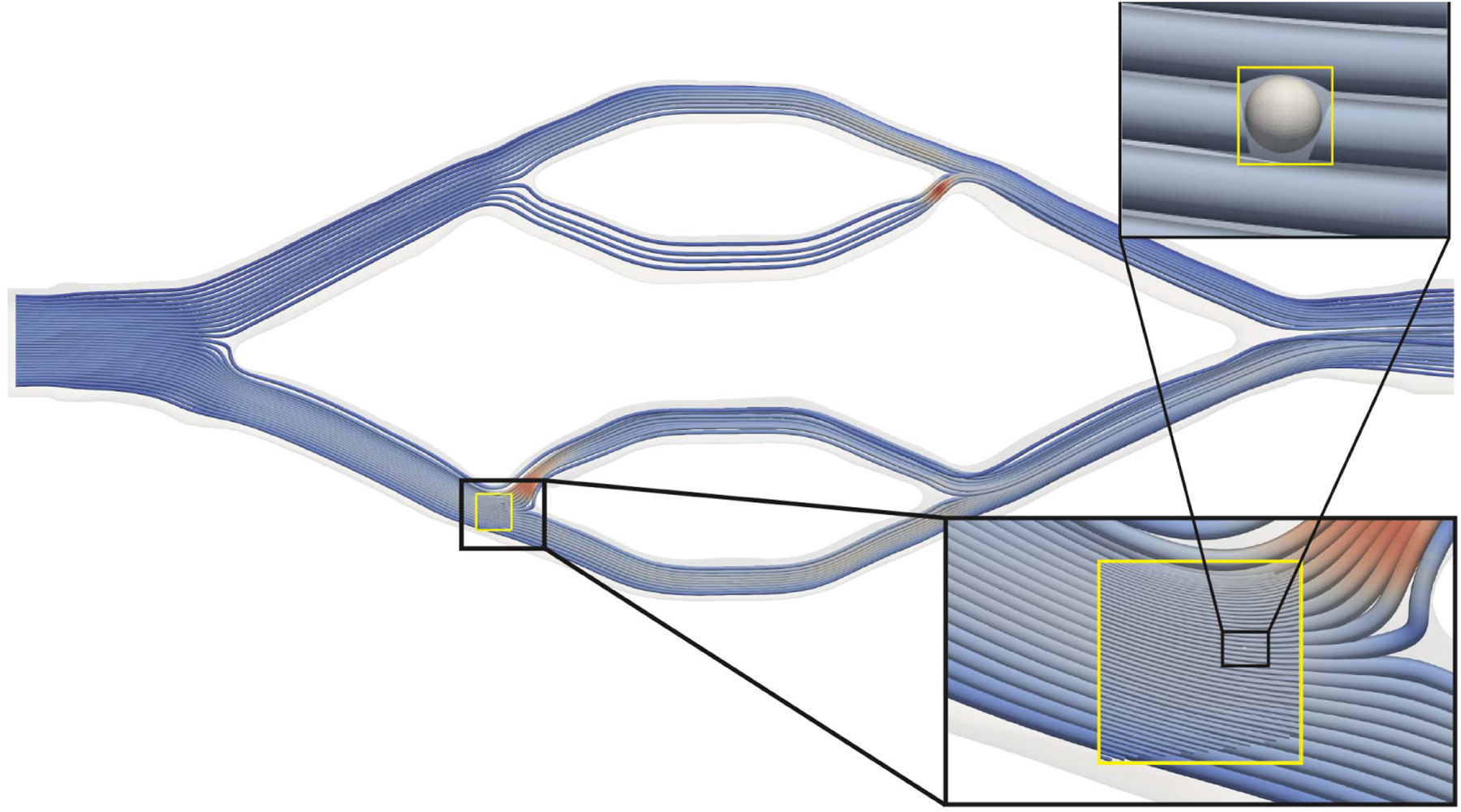
Fig. 9.

Simulation of a large, asymmetric bioprinted vascular bed (via [40]) using the APR algorithm. Streamlines in the window at 4.0 × 10⁵ fine time steps, corresponding to 8.0 × 10⁴ coarse time steps in the bulk are visualized. The cell is also presented at the same time—which is when it was inserted into the simulation. The two insets progressively zoom to the simulated cell. The top-right inset shows the simulated cell with a cut-away from the nearby streamlines.
