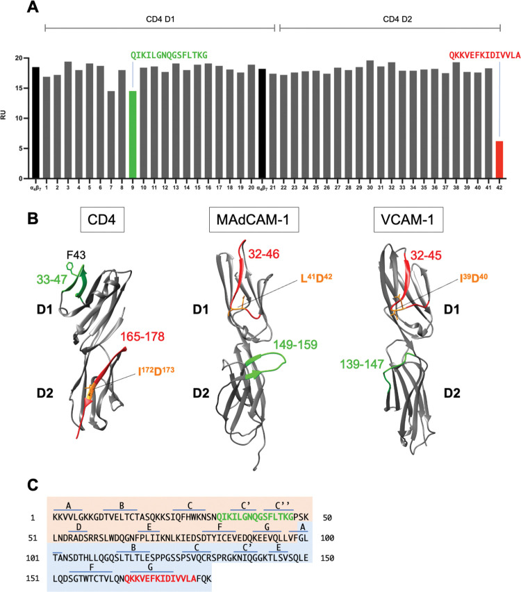
Fig 4. CD4 peptide inhibition of soluble ⍺4β7 binding to CD4 D1D2.
(A) SPR assay of soluble ⍺4β7 binding to a biosensor surface coated with sCD4 D1D2 in the absence (black) or presence of overlapping 15 aa peptides corresponding to residues encoded in CD4 D1 (p1-p21) and D2 (p22-p42). Binding of ⍺4β7 to the D1D2 coated surface is reported in response units (RU). p9 (green) and p42 (red) aa sequences are reported. (B) Ribbon diagram of D1 and D2 domains of CD4 (PDB: 1CDI), MAdCAM-1 (PDB: 1BQS) and VCAM-1 (PDB: 1IJ9). 1° binding sites are highlighted in red, and accessory binding sites are highlighted in green. Predicted metal ion coordination sites for each ligand are indicated (orange). (C) CD4 D1D2 residues with β-strand designations listed above. D1 residues are highlighted in orange and D2 residues in blue. p9 and p42 are highlighted in red and green.

