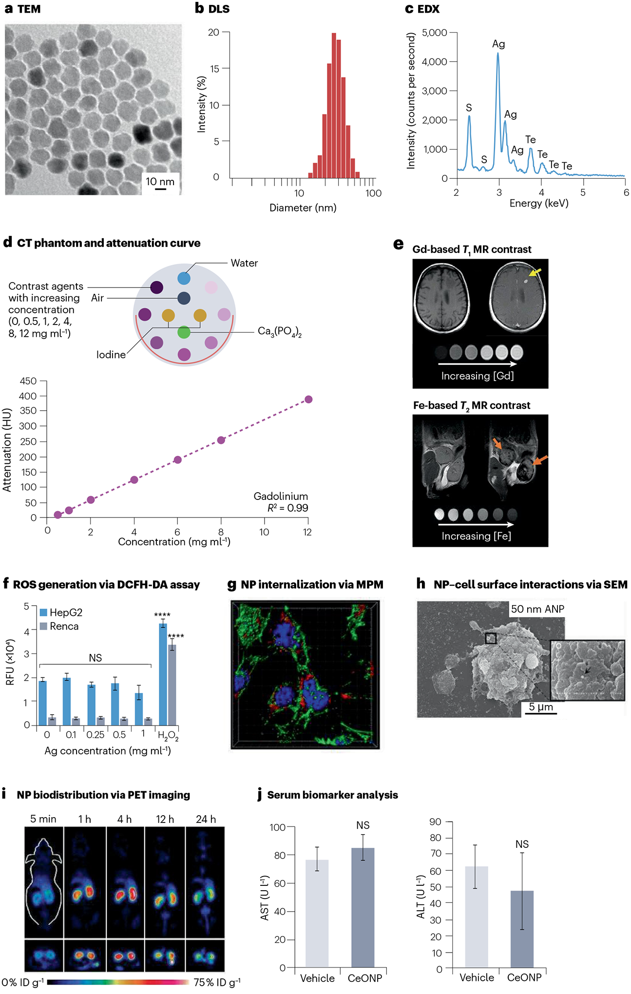
Fig. 3 |. Examples of physical characterization, imaging assessments and biological interactions of nanomaterial-based contrast agents.

a, Transmission electron microscopy (TEM) assesses the nanoparticle (NP) core diameter. b, Dynamic light scattering (DLS) measures the NP hydrodynamic diameter. c, Energy-dispersive X-ray spectroscopy (EDX) identifies the elemental composition of NP. d, Example of a computed tomography (CT) phantom (inset) and attenuation curve derived from the corresponding CT phantom images. e, NPs based on gadolinium (Gd) and iron (Fe) are commonly used as T1-weighted and T2-weighted contrast agents for magnetic resonance imaging (MRI), respectively. Yellow arrow indicates brightening of a brain metastasis due to Gd via T1-weighted MRI. Orange arrows indicate darkening of mammary gland tumours due to Fe via T2-weighted MRI. f, In vitro intracellular reactive oxygen species (ROS) generation is commonly investigated by the 2′,7′-dichlorofluorescein diacetate (DCFH-DA) assay. Data are presented as mean ± s.d. ****P < 0.0001. g, Subcellular resolution multiphoton microscopy (MPM) evaluates cellular internalization of the NP. h, Scanning electron microscopy (SEM) visualizes the interactions between the NP and cell surface. i, In vivo biodistribution (percentage of injected dose per gram of tissue, %ID g−1) is easily determined using positron emission tomography (PET) imaging with radiolabelled NPs. j, Serum biomarkers from blood samples indicate potential NP toxicity and organ damages, such as alanine transaminase (ALT) and blood urea nitrogen (BUN) for liver and kidney functions, respectively. AST, aspartate transferase; CeONP, ceriumoxide nanoparticles; HepG2, hepatocellular carcinoma; NS, not significant; Renca, renal cell carcinoma; RFU, relative fluorescence units. Parts a and b reprinted from ref. 91, Springer Nature Limited. Parts c and f reprinted with permission from ref. 5. Copyright 2022 American Chemical Society. Part d reprinted from ref. 110, Springer Nature Limited. Part e reprinted with permission from ref. 112, Wiley. Part g reprinted from ref. 123, Springer Nature Limited. Part h reprinted from ref. 124, Springer Nature Limited. Part i reprinted from ref. 125, CC BY 4.0. Part j reprinted with permission from ref. 64. Copyright 2021 American Chemical Society.
